
Written by Anh Duong
During June and July of 2020, I conducted research at Dr. Fadok’s lab under the supervision of post-doctoral fellow Maria Dorofeikova. The objectives of this research were to perform a topographic mapping of the basal forebrain cholinergic neurons and their projections to the medial prefrontal cortex (mPFC) by using retrogradely traveling fluorescent beads injected in the mPFC; to perform immunohistochemistry (IHC) staining in cholinergic neurons of the basal forebrain; and to perform anterograde tracing with viral vector Cre-dependently expressing in cholinergic neurons. Since the loss of basal forebrain cholinergic neurons and their cortical projections to areas such as the Prefrontal cortex are associated with Alzheimer Disease (Hampel et al., 2018) and dementia with Lewy bodies (Grothe et al., 2014), the topographic map can provide advancements in understanding the causes and effects of neurodegeneration in dementia and Alzheimer’s disease.
In the first phase of research, Dr. Dorofeikova injected viral vectors (pAAV-hSyn-DIO-EYFP AAV5) and fluorescent latex red beads (Lumafluor) into the mice’ brains. After 3 weeks after the injections, transcranial perfusions were performed to extract nervous tissue for analysis. After observing Dr. Dorofeikova and lab members perform perfusions, I got the chance to practice and do perfusions on my own for the first time during my summer research experience. After the brains were perfused with 4% formalin, I collected 80 nm slices of tissue from where the caudoputamen appears until where the hippocampus begins (where the basal forebrain is located). Every third slice was chosen for immunohistochemistry. Under the guidance of lab technician Alexis Resendez, I performed cholinergic cell staining (with ChAT – Choline Acetyltransferase – antibody) and mounted the stained slices for imaging. I was shown how to use the confocal microscope to assess the area with fluorescent markers that is encoded by the virus and the beads and determine the area of the diagonal band (structure within the basal forebrain) that shows both fluorescent markers. In the pictures, neurons with orange fluorescent markers represent cholinergic cells, red fluorescence shows the beads which travelled from mPFC and represent the innervation between the basal forebrain and the mPFC. cells in the diagonal band that have both orange and red fluorescent shows the cholinergic innervation between the basal forebrain and the mPFC. We extended the research project by observing the GABA-ergic pathways between the basal forebrain and the mPFC (green fluorescent markers are the pAAV-hSyn-DIO-EYFP viral vectors that Cre-dependently expressed in GABA-ergic neurons). We picked 7 slices from 2 mice that clearly show these neurons and found that 82.6% (38 / 46*) of neurons projecting from diagonal band to infralimbic part of prefrontal cortex are cholinergic, while 17.4% are GABA-ergic. The association between apoptosis of cholinergic neurons of diagonal band and cognitive deficits is confirmed through Dr. Dorofeikova’s behavior experiments that show how ablation of cholinergic neurons leads to cognitive deterioration, especially impaired attention and motivation. We intend to continue this research by studying the cFos gene expression (a transcription factor that is thought to mediate long-term changes in neural functioning) after cognitive tasks to elucidate which brain structures are implicated in which cognitive functions, and continue to look at the link between cholinergic cell loss and cognitive deficits seen in dementias.
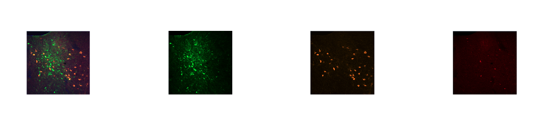
Pictured above: Orange: cholinergic neurons; Red: neurons traveling from basal forebrain to mPFC; Green: GABA-ergic neurons
I am very grateful that the Newcomb-Tulane College has granted me funding for my living expenses, so that I could stay in New Orleans to fulfill my summer research experience. I want to thank the sponsors for allowing me to enrich my educational experience at Tulane through this program. The summer research opportunities have helped me learn new laboratory techniques such as perfusions and IHC staining and progress rapidly in the lab. The hands-on experience has advanced my understanding in neuroscience, particularly in cholinergic neurons and guided me towards developing an elaborate research plan about neurodegeneration diseases for my Honors Thesis.
